Anatomy
The knee joint is made up by 3 separate bones and is the largest joint in humans:
-
The bottom end of the femur – (thigh bone)
-
The top end of the tibia (shin bone)
-
The back of the patella (kneecap)
These 3 bones make up the 3 compartments of the knee:
-
Medial (inner) Tibiofemoral compartments - between the femur and the tibia
-
Lateral (outer) Tibiofemoral compartment - between the femur and the tibia
-
Patellofemoral compartment – between the femur and the patella
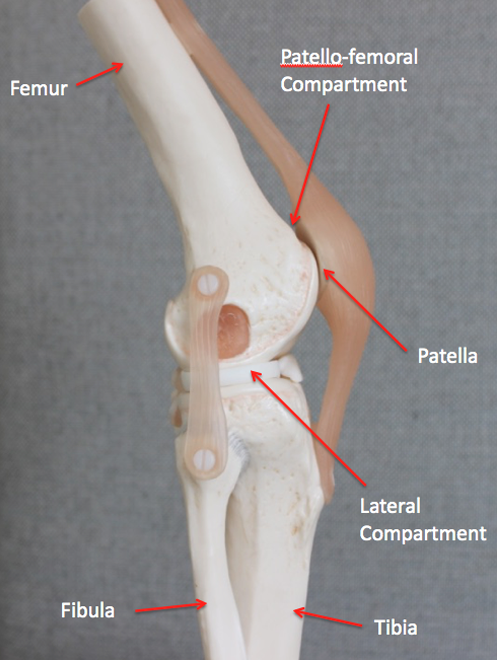 Lateral Side
(outside)
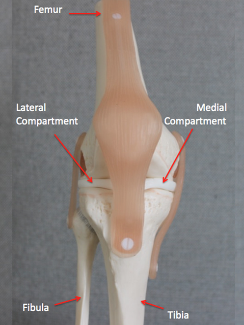 AP
AP
(front)
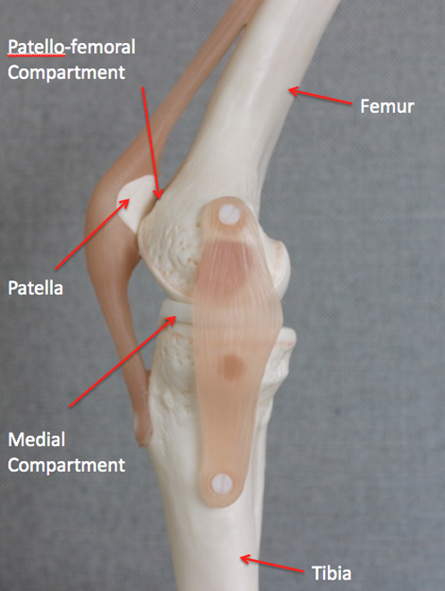 Medial Side
(inside)
Function
The knee acts as a hinge through the complex interaction between the bones, ligaments, muscles and tendons of the joint.
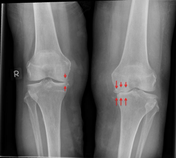
Osteoarthritis (OA)
The surface of each bone of the joint is covered by articular cartilage which allows the bones of the knee to glide over each other during movement. Osteoarthritis occurs due to softening and disintegration of the articular cartilage and is accompanied by new both growth (osteophytic spurs). Osteoarthritis can affect any of the 3 compartments of the knee in isolation or all 3 together. Osteoarthritis classically affects middle aged and older people and can initially present with minimal symptoms.
Diagnosis
-
History & Clinical examination
-
Standing X-rays of the knee
-
Further imaging may be required to specifically assess the joint surface in more detail
-
Diagnostic arthroscopic (keyhole) surgery to assess suitability for certain further procedures may be required
Treatment
Treatment options for OA depend on individual factors:
1) Non operative treatment - This may include weight reduction, activity modification, physiotherapy, the use of non steroidal anti-inflammatories (NSAIDs) and injection therapy
2) Operative treatment – In cases where non-operative treatment has failed or is not appropriate surgery may be required. This can be in the form of: