Anatomy
The hip joint (a ball & socket joint) is made up by 2 bones :
-
The top end of the femur (thigh bone) known as the femoral head (“the ball”)
-
Part of the pelvis called the acetabulum (“the socket”)
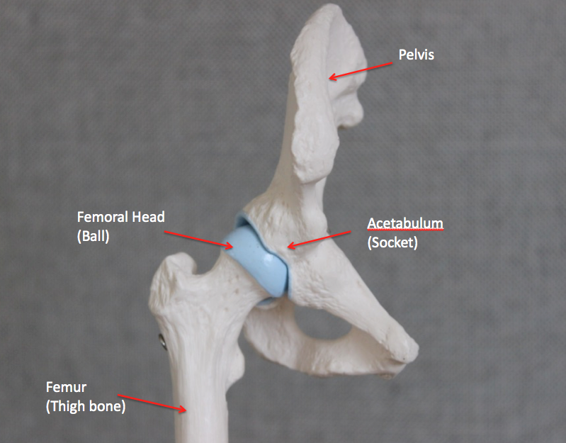
The Hip (right side)
Function
The hip joint connects the leg to the axial skeleton and allows for movements of the leg through the complex interaction between the bones, ligaments, muscles and tendons of the joint.
Osteoarthritis (OA)
The surface of each bone of the joint is covered by articular cartilage which allows the bones of the hip to glide over each other during movement. Osteoarthritis occurs due to softening and disintegration of the articular cartilage and is accompanied by new both growth (osteophytic spurs). Osteoarthritis classically affects middle aged and older people and can initially present with minimal symptoms.
Diagnosis
-
History & Clinical examination
-
X-rays of the pelvis/ hip
-
Further imaging may be required to specifically assess the joint surface in more detail
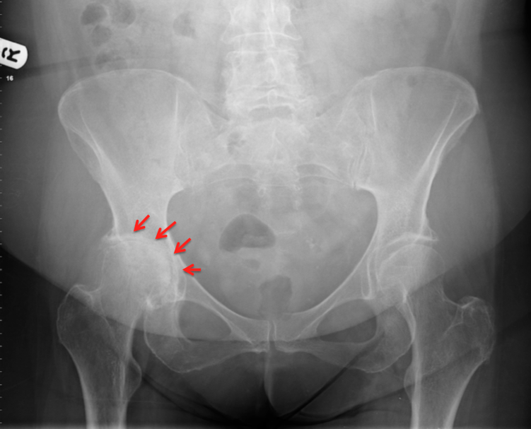
X-ray demonstrating hip osteoarthritis
Treatment
Treatment options for OA depend on individual factors:
-
Non operative treatment - This may include weight reduction, activity modification, physiotherapy, the use of non steroidal anti-inflammatories (NSAIDs) and injection therapy
-
Operative treatment – In cases where non-operative treatment has failed or is not appropriate surgery may be required.
This can be in the form of :
Hip replacement surgery
Robotic assisted total hip replacement